Laser
Safety Questions You Must Ask Before Purchasing a Laser Source
Table of Contents Laser Safety: Risks, Hazards The application of laser is broad. And therefore its impact can be realized everywhere. Today, it is hard
Laser
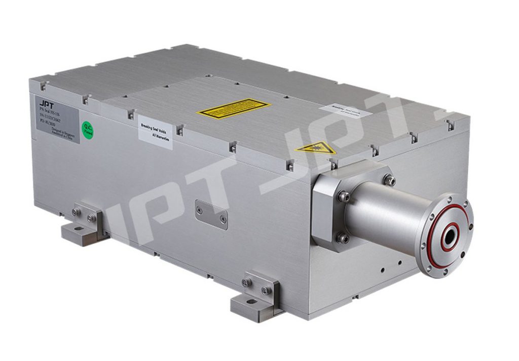
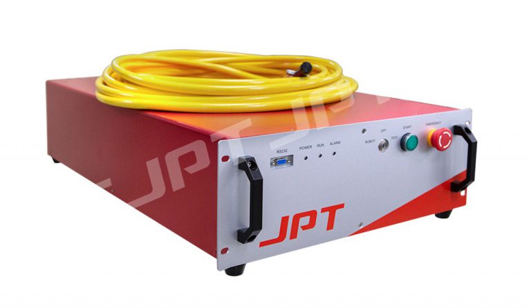
Laser Cleaning: MOPA Pulsed Fiber Laser VS CW Laser
Laser cleaning – using a focused laser beam to rapidly vaporize or strip the contaminants on the material surface. Compare with all kinds of traditional
Writer at JPT Laser
Dr.A.P.Singh is Ph.D. from University of Delhi. He specializes in laser-based materials processing. He was Senior Research Fellow in University of Delhi. He received prestigious JSPS Postdoc fellowship and worked at AIST, Tsukuba, Japan. He has published more than 30 research articles in journals and conferences of international repute. He has about 12 years of experience in academics.
Latest posts by Amit Singh (see all)